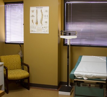
Lhermitte-Duclos disease is a rare lesion characterized by enlarged cerebellar folia containing abnormal ganglion cells. This case report describes a 51-year-old woman who was initially misdiagnosed as having adult-onset aqueductal stenosis. There were no abnormal findings on computerized tomography (CT), but subsequent magnetic resonance (MR) imaging showed a midline cerebellar lesion extending to the brain stem. This is a unique case of Lhermitte-Duclos disease arising within the cerebellar vermis. The characteristic feature of an enlarged cerebellar hemisphere is absent on CT scans; thus MR imaging is needed to confirm the diagnosis. If diagnosed late, this generally benign lesion becomes difficult to resect totally and has a poorer prognosis. Only two reports have mentioned the MR imaging characteristics of Lhermitte-Duclos disease; both described only T2-weighted images. This case illustrates the full spectrum of MR imaging features of this disease. Both T1- and T2-weighted studies showed enlarged cerebellar folia within the lesion. The T1-weighted image showed a mixed iso- and hypodense signal and the T2-weighted image a homogeneously increased signal; with gadolinium administration the lesion did not enhance. The latter feature supports the theory that this disease is a hamartoma rather than a tumor.
Industry Articles
- Dr. Shah Siddiqi Retiring, Texas Spine Center to Close
Dr. Shah Siddiqi, a distinguished neurosurgeon … - Your Guide to Robotic Spine Surgery
In recent years, robotic spine surgery … - Disc Prolapse Recovery Time: How Long Does a Herniated Disc Take to Heal?
Recovering from a disc prolapse, also …