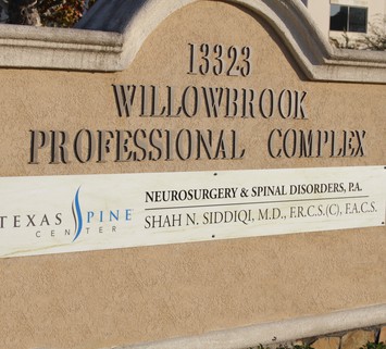
Ventral and Ventrolateral Spine Decompression and Fusion
Please click here to read Dr. Siddiqi's chapter from ‘Spine Surgery: Techniques, Complication Avoidance and Management’, by Ed. E. Benzel.
Please click here to read Dr. Siddiqi's chapter from ‘Spine Surgery: Techniques, Complication Avoidance and Management’, by Ed. E. Benzel.
OBJECTIVE: To determine the effect of initial therapy (surgery and external beam radiation) on the proliferative capacity of glioblastoma and whether adjunctive high focused doses of radiation therapy can further reduce the proliferative capacity of the tumor. This would provide a rationale for attempting to further control local tumor growth with the different forms of high-dose focused radiation available.
We performed a retrospective study of 107 consecutive patients with syndromic forms of craniosynostosis (craniofacial dysostosis) seen by the craniofacial team at the Hospital for Sick Children between 1986 and 1992. There were 51 patients with Crouzon's syndrome, 33 with Apert's syndrome, 8 with Pfeiffer syndrome, 11 with Saethre-Chotzen syndrome, and 4 with kleeblättschadel anomaly. Six patients developed raised intracranial pressure (ICP) after initial suture release and decompression (Apert's syndrome, three patients; Pfeiffer syndrome, one patient; Saethre-Chotzen syndrome, two patients). Raised ICP was considered in those children who returned with a bulging fontanelle, progressive frontal bone protrusion, intermittent headaches, irritability, and vomiting. The diagnosis of raised ICP was based on papilledema (four patients), progressive macrocephaly (one patient), and ICP monitoring (one patient). No child in this group had hydrocephalus requiring cerebrospinal fluid diversion. Once raised ICP was detected in these …
Lhermitte-Duclos disease is a rare lesion characterized by enlarged cerebellar folia containing abnormal ganglion cells. This case report describes a 51-year-old woman who was initially misdiagnosed as having adult-onset aqueductal stenosis. There were no abnormal findings on computerized tomography (CT), but subsequent magnetic resonance (MR) imaging showed a midline cerebellar lesion extending to the brain stem. This is a unique case of Lhermitte-Duclos disease arising within the cerebellar vermis. The characteristic feature of an enlarged cerebellar hemisphere is absent on CT scans; thus MR imaging is needed to confirm the diagnosis. If diagnosed late, this generally benign lesion becomes difficult to resect totally and has a poorer prognosis. Only two reports have mentioned the MR imaging characteristics of Lhermitte-Duclos disease; both described only T2-weighted images. This case illustrates the full spectrum of MR imaging features of this disease. Both T1- …